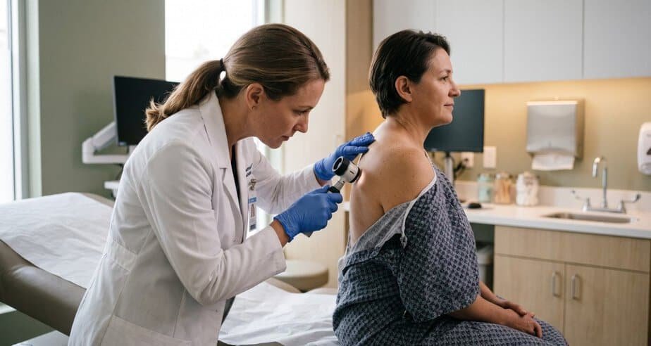
Melanoma can look like a dark mole with ragged edges. It can also look like a pink bump you assume is a stubborn pimple, a sore on your heel that never closes, or a brown stripe under a fingernail you blame on a slammed door. Early melanoma warning signs are easy to explain away, and that is how weeks turn into months.
Skin changes rarely announce themselves loudly. You notice something while drying off after a shower, think “probably nothing,” and forget it by dinner. A spot you filed under “probably nothing” is exactly what a clinician would rather see early.
This list covers six of them, each tied to published research. Some are the classics, like a mole that changes. Others catch the melanomas that slip past the standard checklist. You will also see where the evidence has limits, because a warning sign is only useful when you know how much weight it can carry.
Nothing here replaces a skin exam. Use it to decide what deserves a photo and an appointment.
1. A Mole That Is Changing
Dermatologists call it evolution, and it carries real weight. In 2004, a JAMA review by Abbasi and colleagues recommended expanding the familiar ABCD checklist (asymmetry, border irregularity, color variation, diameter) into ABCDE, with the E standing for evolving. The authors defined change broadly: size, shape, symptoms such as itching or tenderness, surface changes such as bleeding, and shifts in color.
If you want a way to keep track of a spot between appointments, an online photo tool such as ScanSkinAI can apply the ABCDE criteria to an uploaded picture and return a general risk tier. It is a screening aid, not an exam. It cannot diagnose skin cancer or rule it out, and the company’s own page notes it may miss non-pigmented melanomas, so a spot that changes in color, shape, or size still needs a clinician’s eye.
Visible change can show up later than you would hope. A retrospective study of 144 melanomas by Duarte and colleagues found evolution documented in 32.7% of in situ lesions and 50.0% of invasive ones. That gap hints that waiting for obvious change can mean waiting past the earliest stage.
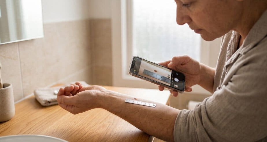
Photograph any mole you are unsure about, with a ruler or coin beside it, and compare the picture a month later. Itching or bleeding in a mole you have carried for years justifies booking an appointment now. Even a quick phone note counts: the date, where the spot sits on your body, and what changed.
2. The Mole That Does Not Match the Others
Your moles tend to resemble one another in color, size, and shape. A melanoma often breaks that pattern, and dermatologists call the outlier the ugly duckling sign.
Scope and colleagues tested the idea in 2008. Thirty-four observers, from pigmented lesion experts to nonclinical medical staff, reviewed photographs of 12 patients who each had at least eight atypical moles. All five melanomas in the set stood out as different to at least two-thirds of the observers, and only 3 of 140 benign lesions did. Sensitivity was 0.90 across the whole group and 0.85 among the nonclinical staff.
The limits matter. The study used photographs instead of live skin exams, and every patient already had many unusual moles, so the numbers will not transfer neatly to a casual self-check. The authors also suspected that seborrheic keratoses, a benign growth, could fool the method.
Stand in front of a mirror and ask which mole looks least like its neighbors. That one gets shown to a clinician. Your back is the hardest area to judge because you cannot see it, so ask a partner or friend to photograph it, or have a clinician do the comparison.
3. Uneven Shape, Ragged Borders, Patchy Color
The ABCDE checklist is the one most people have heard of, and it still earns its place. Its authors stressed that it was built to help nondermatologists tell common moles from cancer, and that it does not capture every melanoma feature.
Look for halves that do not match, an edge that looks scalloped or smudged, and several shades inside one mole. The 6 mm diameter cutoff, roughly the width of a pencil eraser, gets the most attention. The 2004 review examined small melanomas and concluded the data did not support lowering that cutoff, yet it still put change over time at the center of its advice. Size alone should never settle the question.
Duarte’s team found that the more ABCDE signs a lesion carried, the likelier it was that someone other than a dermatologist suspected melanoma first, ranging from 8% with no signs to 32% with all five. They also concluded that relying on the rule by itself might miss early melanomas. Treat a lack of ABCDE signs as limited reassurance.
4. A Firm, Raised Bump That Keeps Growing
Nodular melanoma grows downward into the skin from early on, which means it can skip the flat, irregular look the checklist expects. In a 2003 questionnaire study of 125 patients at Melbourne’s Victorian Melanoma Service, Chamberlain and colleagues reported that nodular melanomas were more often symmetric, raised, uniform in color, and nonpigmented than the superficial spreading type.
That profile explains the EFG rule: elevated, firm, growing. A new bump that feels solid, rises above the skin, and keeps enlarging deserves a prompt exam even when it is one even color, pink, or the same shade as your skin.
Plenty of harmless lumps are raised and firm too, a dermatofibroma being the usual example. The sign calls for an exam, and the exam can sort it out.
One caution on the evidence: those findings come from patients describing their own lesions, so recall shapes them, and the study cannot tell you how often a given bump turns out to be melanoma.
5. A Pale Or Pink Spot That Will Not Heal
Amelanotic melanoma lacks the dark pigment people associate with the disease, so it blends in with ordinary skin problems. A 2024 systematic review by Cassalia and colleagues gathered 26 cases of acral amelanotic melanoma (palms, soles, and under the nails) that were first diagnosed as something else. The initial labels were non-healing ulcers or injuries in 37.5% of cases, benign growths in 29.2%, and infections in 20.8%. The foot was the most common site at 53.8%.
Biopsy use varied sharply by location. Histological evaluation was done in 50% of upper extremity cases, compared with 7.1% of foot cases and none of the heel cases, and the authors concluded that underuse of biopsy on the lower limb contributes to misdiagnosis. The percentages come from only 26 reported cases, so they illustrate a pattern and carry limited statistical weight.
A sore that keeps failing to heal, a pink or red bump that keeps growing, or a wart-like patch that ignores treatment should be seen by a clinician. Asking whether a biopsy makes sense is a fair question.
6. A Dark Streak in A Nail
A pigmented band running the length of a nail, called longitudinal melanonychia, is common and usually harmless. In people with darker skin tones it often appears on several nails at once. A band on a single nail in an adult raises more suspicion.
Edwards, Usatine, and Heath, writing in Cutis in 2024, described irregularly spaced brown-to-black lines of varying width as characteristic of subungual melanoma. Pigment spreading onto the skin beside the nail, known as Hutchinson sign, is another worrying feature. They also cautioned that a history of injury can mislead, because nail changes blamed on trauma may turn out to be melanoma. Subungual melanoma tends to be found late, and people of African and Asian descent and American Indian individuals face the worst outcomes, largely because of advanced stage at diagnosis.
Photograph the nail every few weeks. The same authors encouraged photos because change over time guides the decision to biopsy, and a nail matrix biopsy is the diagnostic standard when cancer is suspected.
The Six Signs at A Glance
| Red flag | What you may see | What the research adds |
| Changing mole | Growth, new shape, itching, tenderness, bleeding, color shift | Evolution was added to ABCD in a 2004 JAMA review (Abbasi et al.) |
| Odd one out | One mole unlike the rest | All 5 melanomas in a 2008 observer study stood out (Scope et al.) |
| Uneven appearance | Lopsided shape, ragged border, mixed shades | ABCDE alone may miss early lesions (Duarte et al., 2021) |
| Firm, raised bump | Dome-shaped, one color or pink, keeps growing | Nodular type is more often symmetric and nonpigmented (Chamberlain et al., 2003) |
| Pale, non-healing spot | Sore or bump on palm, sole, or nail area | First labeled ulcer, benign growth, or infection in most reported cases (Cassalia et al., 2024) |
| Nail streak | Irregular dark band, pigment on nearby skin | Single nail in an adult warrants closer attention (Edwards et al., 2024) |
What to Do This Week
Pick one evening. Undress in good light, photograph any mole, bump, sore, or nail band that bothers you with a ruler beside it, and set a reminder to compare in a month. Anything that itches, bleeds, grows, or looks different from its neighbors goes on the appointment list.
At the visit, name the sign that worried you, ask whether dermoscopy or a biopsy is warranted, and ask for your other moles to be examined too, since the ugly duckling comparison depends on seeing the whole picture.
This article is educational and does not diagnose any condition. Only a clinician who examines your skin can do that.
Reade next: Building a Practical Skin Care Plan with Local Dermatology Support
References
Abbasi, N. R., Shaw, H. M., Rigel, D. S., Friedman, R. J., McCarthy, W. H., Osman, I., Kopf, A. W., & Polsky, D. (2004). Early diagnosis of cutaneous melanoma: Revisiting the ABCD criteria. JAMA, 292(22), 2771-2776. https://doi.org/10.1001/jama.292.22.2771
Cassalia, F., Danese, A., Cocchi, E., Danese, E., Ambrogio, F., Cazzato, G., Mazza, M., Zambello, A., Belloni Fortina, A., & Melandri, D. (2024). Misdiagnosis and clinical insights into acral amelanotic melanoma: A systematic review. Journal of Personalized Medicine, 14(5), 518. https://doi.org/10.3390/jpm14050518
Chamberlain, A. J., Fritschi, L., & Kelly, J. W. (2003). Nodular melanoma: Patients’ perceptions of presenting features and implications for earlier detection. Journal of the American Academy of Dermatology, 48(5), 694-701. https://doi.org/10.1067/mjd.2003.216
Duarte, A. F., Sousa-Pinto, B., Azevedo, L. F., Barros, A. M., Puig, S., Malvehy, J., Haneke, E., & Correia, O. (2021). Clinical ABCDE rule for early melanoma detection. European Journal of Dermatology, 31(6), 771-778. https://doi.org/10.1684/ejd.2021.4171
Edwards, T. M., Usatine, R. P., & Heath, C. R. (2024). Longitudinal melanonychia. Cutis, 113(3), 143-144. https://doi.org/10.12788/cutis.0972
Scope, A., Dusza, S. W., Halpern, A. C., Rabinovitz, H., Braun, R. P., Zalaudek, I., Argenziano, G., & Marghoob, A. A. (2008). The “ugly duckling” sign: Agreement between observers. Archives of Dermatology, 144(1), 58-64. https://doi.org/10.1001/archderm.2007.15